Functional Neuroimaging
-
Group Leader
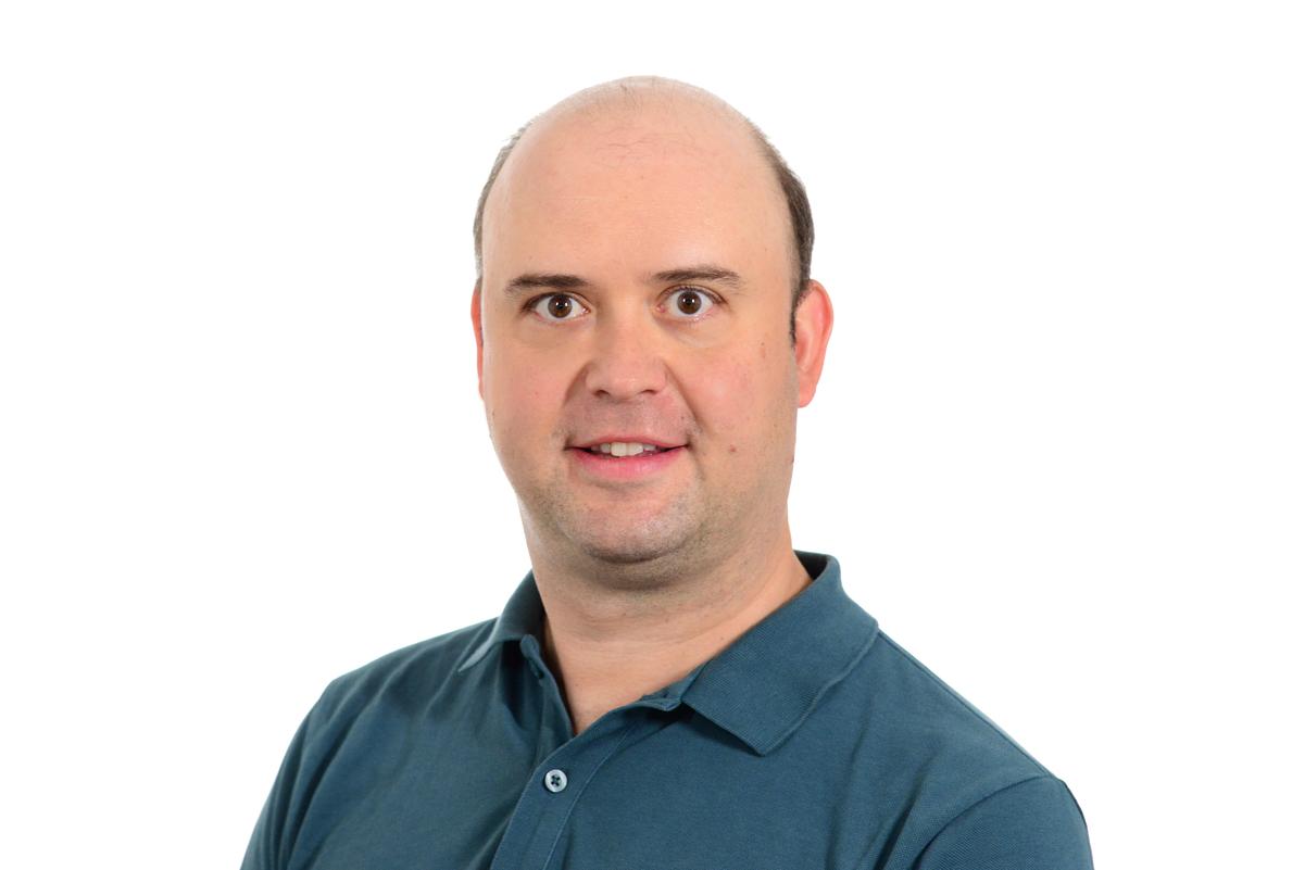
Prof. Dr. med. Sophia Stöcklein
Senior physician, head of MRI, head of research organisation
Sophia has been involved in advanced neuroimaging, especially functional connectimocs ever since her doctoral thesis, where she worked on changes of the default network in mild cognitive impairment and Alzheimer’s disease. She went on to gain profound expertise and skills in computational neuroscience and cognitive psychology during a 2-year, DFG-funded postdoctoral fellowship in computational neuroscience at the Martinos Center of Biomedical Imaging (MGH) and the Department of Psychology at Harvard University (Cambridge, USA). Her postdoctoral work focused on the assessment of the reliability and variability of fMRI-based functional connectivity measures and on the explorations of individual differences in the connectivity architecture of the human brain. During her residency training in radiology she went on to apply refined techniques of personalized fMRI analyses to patient cohorts including neuro-degenerative and neuro-oncologic diseases. The overarching aim of her research work is to develop and validate fMRI-based biomarkers that inform personalized diagnostic and therapeutic strategies in the management of patients with neurological disorders.Clinical and Postdoctoral Researchers

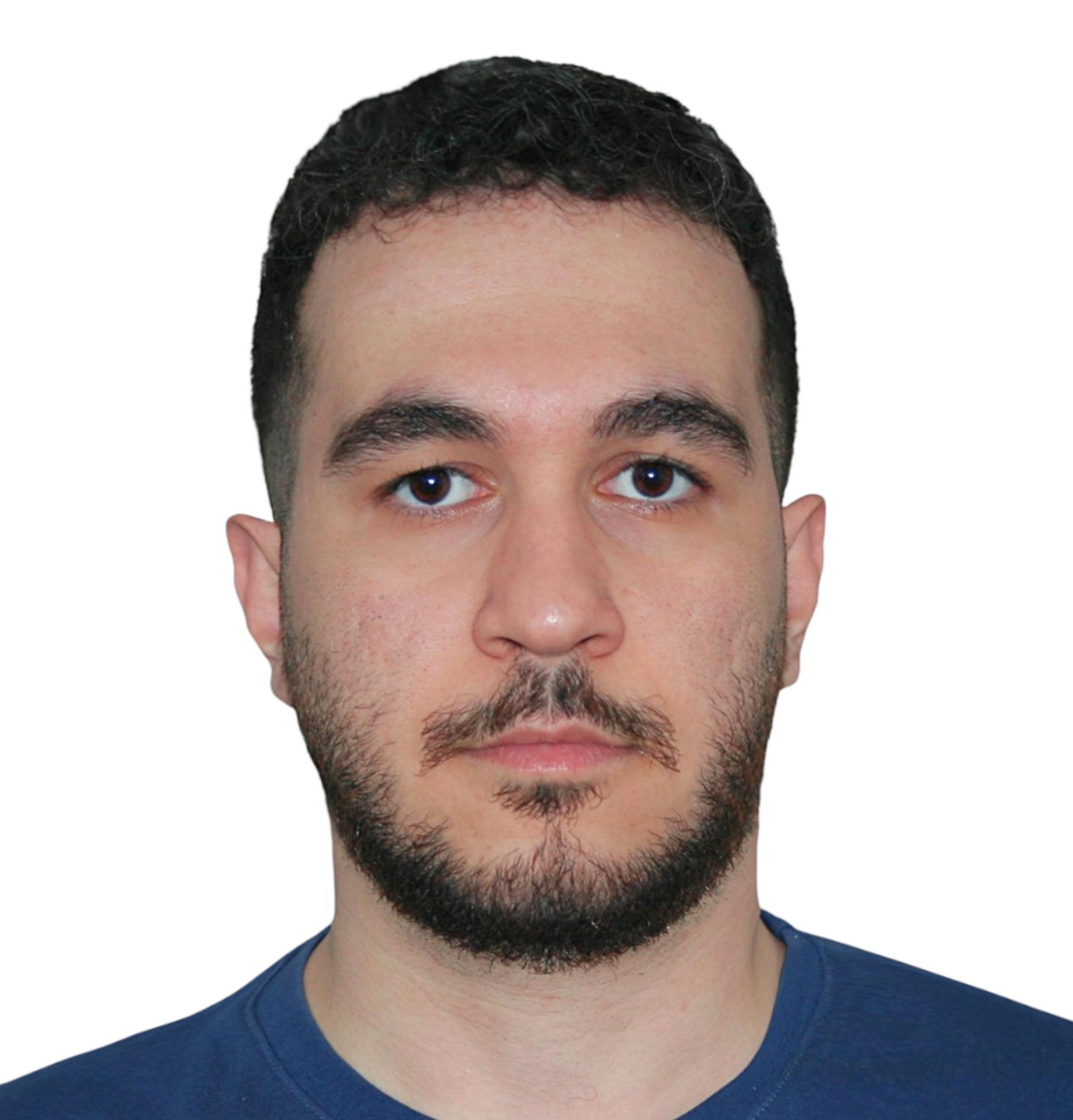
Dr. med. Gloria Biechele
Resident
Gloria received her awarded MD in the field of preclinical molecular brain imaging in mouse models of neurodegenerative disease. Building on this foundation, she is currently translating and expanding these methods to the clinical setting by implementing advanced quantitative MRI techniques on the individual patient level in Alzheimer’s disease, focusing on the interface of functional and quantitative neuroimaging. This approach aims to inform early diagnosis and support the stratification of personalized treatment strategies, particularly in light of emerging therapeutic options. In parallel, she maintains a strong clinical focus in fetal MRI, with postdoctoral research dedicated to the application and advancement of fetal cardiac MRI.Xäüplg-AlSiyziäiJvim-ful#vfiuyziusmi
Franziska Müller, M.Sc.
Resident
Franziska is a radiology resident who also holds a M.Sc. in Human Biology. Her focus has been on systems neuroscience and neuroimaging in both basic and clinical research. She is currently working on functional and structural MRI, and clinical applications of AI solutions in neuroimaging.Äpgußlcog-SOfiääipvim ful_vfiuyziu/mi
Dr. med. Sarah Schläger
Resident
Sarah is a resident and clinician scientist. Her research focuses on novel MRI techniques and integrating AI in the MRI workflow. Currently, she explores innovative MRI applications, such as quantitative imaging in neuromuscular diseases and advanced sequence techniques in fetal MRI.RDgpgz/Ryzägnixipvimefnul_vfiuyaziuemiStefan Suvak
Stefan is a radiology resident with a previous research focus and passion for neurofunctional imaging. As part of his doctoral thesis at the Klinikum rechts der Isar, he investigated the growth behavior of gliomas and the effects of radiotherapy on the functional status of patients in terms of motor or language deficits. Cortical functional imaging using transcranial magnetic stimulation (TMS) was combined with DTI via fiber tracking and correlated with radiotherapy.
RbiwgueRfqgovWim-fulrnvfiuyziuenmi
Stephan Wunderlich, M.Sc.
With a multidisciplinary background in medicine and engineering from TUM, the Martinos Center for Biomedical Imaging in Boston, the Brain Science Center in Edinburgh, and the University of British Columbia, Stephan’s research passion lies at the intersection of technical methodologies and medicine. He specializes in health data analytics and functional neuroimaging using artificial intelligence methods.
Rbiözgu aUfumipälyz:vim fulrvDfiuySziu miPhD and MD students

Artur Toloknieiev
A doctoral medical student and programmer autodidact, Artur brings to the table a profound understanding of the physiology behind the clinical pictures and a wealth of skill in software engineering with a focus on artificial intelligence. He works on machine learning and data operations, communicates with engineers on medical aspects of the technology and works on the optimization data acquisition, data enrichment and model training processes.
Küäüouliliq Fpbfpygvöfc/ävf-mi
Tobias Prester
Tobias successfully completed his medical studies at the LMU and is currently working on his medical doctor’s degree in the field of fetal MR imaging. His doctoral research project focuses on the effects of a SARS-CoV-2 infection during pregnancy on the fetus and the placenta.
Junlin Jing
Junlin is a Ph.D. student whose previous research focused on machine learning and the application of ICA-derived approaches to functional neuroimaging and structural connectivity. He is currently working on the use of machine learning to investigate abnormal functional connectomes of neurological and psychiatric disorders, particularly in personalized diagnosis.
QfuäluYsQluxvim ful_vfiuyWziu YmiUndergraduate students

Hlib Kholodkov
A computer science specialist and model development enthusiast, Hlib combines expertise in data preprocessing, scientific analysis, and machine learning. He focuses on optimizing model workflows, from data acquisition and enrichment to training, while bridging technical and theoretical insights.
Dmytro Voitsekhivskyi
Dmytro uses machine learning and imaging algorithms to advance computational neuroscience. He holds a degree in Computer Science from the Technical University of Munich and is currently pursuing a degree in Management & Digital Technologies at the Ludwig Maximilian University of Munich. His research efforts include data preprocessing, deep learning model development, and the creation of high-performance computing solutions aimed at improving neuroimaging techniques through cutting-edge AI innovations.

Roman Lvovich
A student of Electrical Engineering and Information Technology at the Technical University of Munich, Roman has a solid background in mathematical methods, computational techniques, and simulation of complex systems. He is currently focusing on the mathematical aspects of AI applications in neuroscience, including multidimensional data analysis, modeling of neural processes, and development of algorithms for interpretation of complex neural network dynamics.

Mohammad Saeed Motevali
A computer science student specializing in medical image processing and analysis. His research focuses on developing algorithms for processing and analyzing fMRI data, with a particular emphasis on machine learning applications in medical diagnostics. His work combines expertise in computational methods with practical applications in abnormality detection, aiming to improve diagnostic capabilities through automated analysis of neural imaging data. He is particularly interested in developing robust machine learning and deep learning frameworks for processing complex neurological data and identifying patterns that can aid in the early detection of neurological conditions through fMRI analysis.
Technicians

Boris Papazov
Boris received his Bachelor of Science in the field of Engineering Physics and his Master of Science in the field of Aerospace Engineering Sciences. He currently organizes, optimizes, and performs clinical research MRI scans with a focus on fMRI and brain tumors.
Scientific Project Management

PD Dr. Enrico Schulz
Enrico is a psychologist by training and provides scientific and administrative support to the team.
Dr. rer. nat. Julia Ruat
Julia is a neurobiologist by training and provides scientific and administrative support to the team.
-
The Functional Neuroimaging Group focuses on the assessment of the “individual brain connectome” based on resting-state functional magnetic resonance imaging (fMRI). We work on algorithms to detect and quantify changes in the functional connectivity architecture of individual patients in comparison to large reference cohorts, with the overarching aim to develop imaging biomarkers for diagnosis, treatment monitoring and therapy guidance of neurological and neuropsychiatric disorders. Our main focus comprises neuro-oncological and neuro-degenerative diseases. In order to better understand the adult brain, our group further uses advanced MRI techniques to study brain development perinatally, including fetal and neonatal imaging of the brain, and organ systems that impact brain development such as the cardio-respiratory system.
Our interdisciplinary group is composed of radiologists, neurobiologists, psychologist, engineers, and computer scientists.
Functional connectivity in neurological disorders
New concepts of brain disease suggest that network disruption does not only occur as an epi-phenomenon of underlying brain pathology, but that the formation of aberrant connections can be a major contributor to the progression of brain diseases, including neuro-oncological and neurodegenerative diseases. The current role of MRI in such diseases is to provide structural information about tumors and their close vicinity and to describe patterns of brain atrophy and vascular damage. Here, assessment of functional connectivity alterations on the whole brain level might provide important additional information that is not yet available in vivo in individual patients.
However, despite more than two decades of functional connectivity research no clinically applicable and validated imaging tool is available to assess network dysfunctions quantitatively in the individual patient. We have designed a novel imaging marker based on resting-state fMRI which assesses global functional connectivity in individual patients (Stoecklein et al., 2020), with the aim to detect and quantify alterations of the connectivity architecture of individual patients by comparing individual connectivity profiles to a normative distribution derived from large, healthy reference cohorts.
Neuro-oncology
Building on our long-standing work on patient-level functional connectivity mapping, our group is currently advancing these concepts toward clinical translation in neuro-oncology through the ERC Consolidator project CONNECT.
CONNECT - an ERC Consolidator project

Recent advances in cancer neuroscience show that malignant brain tumors such as glioblastoma extend far beyond the visible lesion and interact with brain-wide neuronal circuits. By forming synaptic connections with neurons, tumors can exploit neural activity to support their growth, strongly influencing disease progression.
With the ERC Consolidator project CONNECT (Cutting-Edge Neuroimaging for Functional Brain Network Evaluation in Cancer Patients), we aim to translate these insights into clinical imaging by establishing functional connectivity MRI as a quantitative tool for cancer diagnostics.
Central to the project is the construction of a large-scale reference database comprising thousands of fMRI datasets. Leveraging this resource, we will develop AI-supported methods to map and quantify patient-specific network deviations. These imaging findings will be linked to structural connectivity and molecular tumor profiles, enabling mechanistic insight into tumor–brain interactions.
The clinical potential of this approach will be evaluated in two independent patient cohorts. By identifying network changes that may precede visible tumor progression, CONNECT aims to enable earlier detection of recurrence or metastasis and to inform more precise, network-oriented therapeutic strategies.
Neurodegeneration
Recent advances in functional neuroimaging highlighted disruptions in brain network in neurodegenerative disorders such as Alzheimer’s Disease, and Parkinson’s Disease. We aim to develop a functional imaging marker that predicts disease progression and assesses treatment response. Recent publications suggest the propagation of neurodegenerative pathology, e.g., tau spread, through functional brain network disruptions.
Artificial intelligence for data analysis of large cohort studies
In radiomics research, artificial intelligence (AI) has emerged as a powerful tool to efficiently handle large multidimensional datasets. In particular, graph auto-encoders have proven to be potent instruments for unsupervised anomaly detection. We aim to develop AI algorithms that detect abnormalities in functional connectivity not only based on 4D functional MRI data, but also based on enriched multidimensional input data including demographics (age, sex, gender) and technical parameters (scanner type and protocol specifications). AI-based abnormality detection has been applied to structural MRI data, however moving this field into the fMRI arena and leveraging large publicly available databases as reference samples offers great potential for developing a refined diagnostic tool that is perfectly tailored to each individual patient.Perinatal imaging: Advanced MR imaging for the exploration of brain development in health and disease
To understand the structure and function of the adult brain, it is crucial to understand its earliest development, including potential disruptive factors. We investigate brain development in utero and in the perinatal period, focusing on physiological development but also on early pathology of brain development, and how it might be affected by other organs such as the cardiorespiratory system.
-
ALZECA
Alzheimer's disease is characterized by the presence of amyloid plaques - protein depositions - in the brain. Currently, assessing amyloid presence and distribution in the brain requires positron emission tomography (PET). The aim of the ALZECA study is to determine whether this procedure may be replaced by magnetic resonance imaging (MRI) using the contrast agent ADx-001. ADx-001 is a novel, intravenously delivered, Gd-containing molecularly targeted liposomal product that is being developed for use in contrast-enabled MR imaging of amyloid plaques.
Replacing PET by MRI would reduce radiation exposure and simplify the diagnostic process by providing detailed information on brain structure and volume while simultaneously identifying amyloid depositions. Animal studies have shown that ADx-001 accumulates in brain regions with amyloid plaques and is detectable by MRI (Badachhape et al., 2020). The objective of this Phase 1 trial is to establish safety of ADx-001 in healthy volunteers, and safety and proof of concept in patients with confirmed amyloid plaques in the brain (confirmed by amyloid PET).
Badachhape, A.A., Working, P.K., Srivastava, M. et al. Pre-clinical dose-ranging efficacy, pharmacokinetics, tissue biodistribution, and toxicity of a targeted contrast agent for MRI of amyloid deposition in Alzheimer’s disease. Sci Rep 10, 16185 (2020)
-
Selected publications
Stoecklein S, Wunderlich S, Papazov B, Winkelmann M, Kunz WG, Mueller K, Ernst K, Stoecklein VM, Blumenberg V, Karschnia P, Bücklein VL, Rejeski K, Schmidt C, von Bergwelt-Baildon M, Tonn JC, Ricke J, Liu H, Remi J, Subklewe M, von Baumgarten L, Schoeberl F. Functional connectivity MRI provides an imagingcorrelate for chimeric antigen receptor T-cell-associated neurotoxicity. Neurooncol Adv 2023 Oct 24;5(1).
Stoecklein S*, Koliogiannis V*, Prester T, Kolben T, Jegen M, Hübener C, Hasbargen U, Flemmer A, Dietrich O, Schinner R, Dinkel J, Fink N, Muenchhoff M, Hintz S, Delius M, Mahner S, Ricke J, Hilgendorff A. Effects of SARS-CoV-2 on prenatal lung growth assessed by fetal MRI. Lancet Respir Med 2022 Apr;10(4):e36-e37 *shared first.
Stoecklein VM*, Stoecklein S*, Galiè F, Ren J, Schmutzer M, Unterrainer M, Albert NL, Kreth FW, Thon N, Liebig T, Ertl-Wagner B, Tonn JC, Liu H. Resting-state fMRI Detects Alterations in Whole Brain Connectivity Related to Tumor Biology in Glioma Patients. Neuro Oncol 2020 Sep 29;22(9):1388-1398 *shared first.
Stoecklein S, Hilgendorff A, Li M, Förster K, Flemmer AW, Galiè F, Wunderlich S, Wang D, Stein S, Ehrhardt H, Dietrich O, Zou Q, Zhou S, Ertl-Wagner B, Liu H. Variable functional connectivity architecture of the preterm human brain: Impact of developmental cortical expansion and maturation. Proc Natl Acad Sci U S A 2020 Jan 14;117(2):1201-1206.
Mueller S, Wang D, Fox MD, Yeo BT, Sepulcre J, Sabuncu MR, Shafee R, Lu J, Liu H. Individual variability in functional connectivity architecture of the human brain. Neuron 2013 Feb 6;77(3):586-95.
Full publication list
-
The Functional Neuroimaging group currently receives research funding by

European Research Council

Netzwerk Universitätsmedizin

Medical Faculty of the LMU

Deutsche Gesellschaft für Muskelkranke e.V.
Bruno und Helene Jöster Stiftung
-

Brainlab AG

Deutsches Zentrum für Lungenforschung (DZL)

Deutsches Zentrum für Neurodegenerative Erkrankungen (DZNE)

Helmholtz Zentrum München

Hôpital Necker-Enfants malades, Paris
Medizinische Universität Wien, Abteilung für Neuroradiologie und muskuloskelettale Radiologie

Northh Medical

Institut für Neuropathologie, LMU

Brustzentrum, LMU Klinikum

Institut für Schlaganfall- und Demenzforschung (ISD), LMU Klinikum

Klinik für Psychiatrie und Psychotherapie, LMU Klinikum

Klinik und Poliklinik für Neurologie, LMU Klinikum
 Klinik und Poliklinik für Nuklearmedizin, LMU Klinikum
Medizinische Klinik und Poliklinik V, LMU Klinikum

Neurochirurgische Klinik und Poliklinik, LMU Klinikum


